DLT-Cam Viewer 29_06_21 — программа для камеры
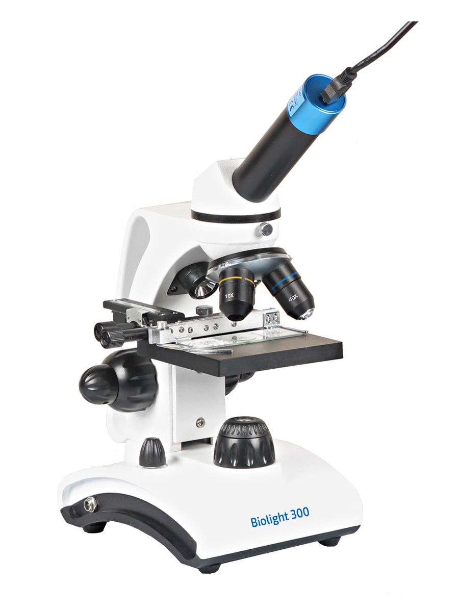
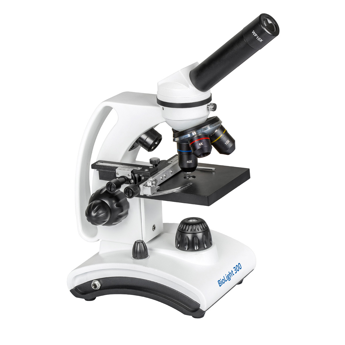
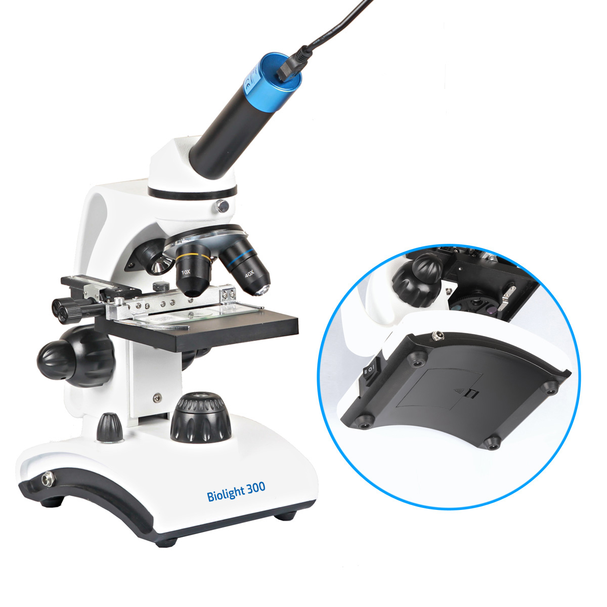
Delta Optical BioLight 300 microscope with the Delta Optical DLT-Cam Basic 2 MP camera
The successor of the most popular biological microscope for young researchers and slightly older micro-world enthusiasts in Poland and Central Europe! The Delta Optical BioLight 300 microscope with a camera is an improved version of the DO BioLight 200 model. BioLight 300 has a completely new design. First of all, the optical system that determines the quality of the microscopic image has been improved. In addition, the microscope has a built-in place for mounting a battery, so that observations can also be carried out without the need to connect to the electrical network. This solution also allows you to take the microscope on a research field trip (e.g. to a pond to observe water microorganisms). Attention is drawn to the modern design of the body, the handle integrated into the tripod makes it easy and safe to carry the microscope (in the case of children, the risk of dropping the device is minimized). The microscope comes with a USB digital camera with the highest resolution in this class of microscopes! The DLT-Cam Basic camera has a resolution of 2 million pixels (that is, it allows you to obtain images with a size of 1600 x 1200 pixels).
Here’s why the Delta Optical BioLight 300 with the DLT-Cam Basic 2 MP camera is the best choice in its class:
- the set includes a digital color camera with a high resolution of 2 million pixels, thanks to which it is possible to save images of very high quality
- English, Russian and other languages extended Delta Optical DLTCamViewer control software included (functions are described below)
- the improved optical system made of the highest quality optical glass allows you to get an even better image — bright and without distortions
- built-in battery operation without the need to connect to a socket — the possibility of taking the microscope into the field
- handle integrated into the tripod for safe carrying of the microscope
- a solid, metal body guarantees long-term reliability, made of light and durable alloys
- double focus adjustment: coaxial macrometric and micrometric knobs — facilitates precise focus adjustment, which is especially important at high magnifications (in this price range it is worth paying attention to it)
- double lighting system with smooth brightness adjustment: passing (lower — so-called DIA) for observing preparations on glass slides (transparent objects, transmitting the light beam) and reflected (upper — so-called EPI). Reflected lighting allows you to observe non-transparent objects (e.g. insects, minerals, fabrics, coins)
- LED lighting prevents the preparation and microscope from heating up (no risk of burns) and guarantees several tens of thousands of hours of operation
- 4x, 10x and 40x achromatic lenses and the WF10x wide field eyepiece
- real range of magnifications obtained thanks to lenses : from 40x to 400x (you can additionally buy a high-quality 16x eyepiece and get a 640x magnification! — see «recommended products»)
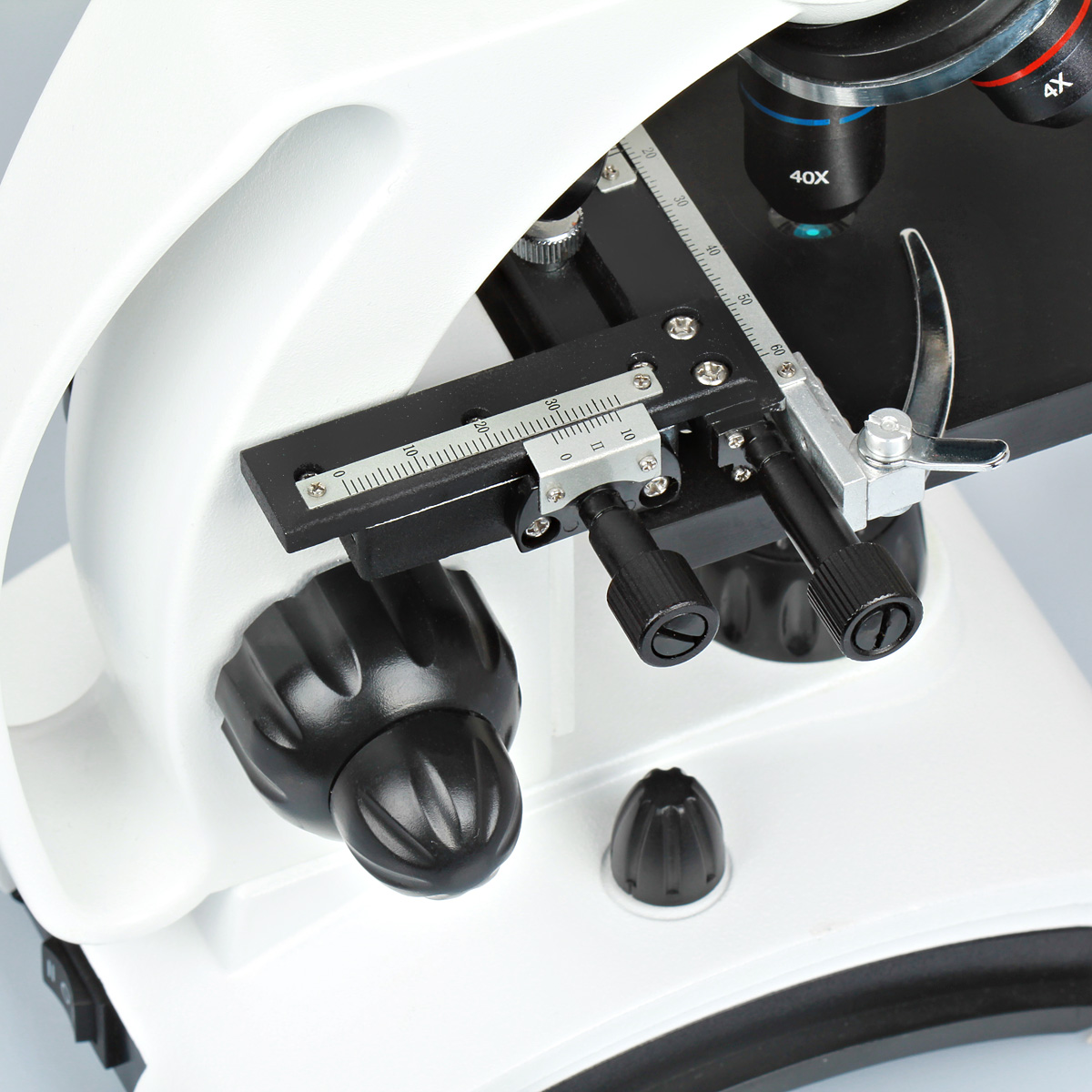
- cross table with specimen holder and precise knobs to move horizontally in the X and Y axes — precise selection of the test area
- the slide mechanism of the preparation has a vernier — a special scale that increases the accuracy of reading
- a six-socket wheel with color filters that allow you to select the optimal lighting conditions and obtain a better image contrast during observation
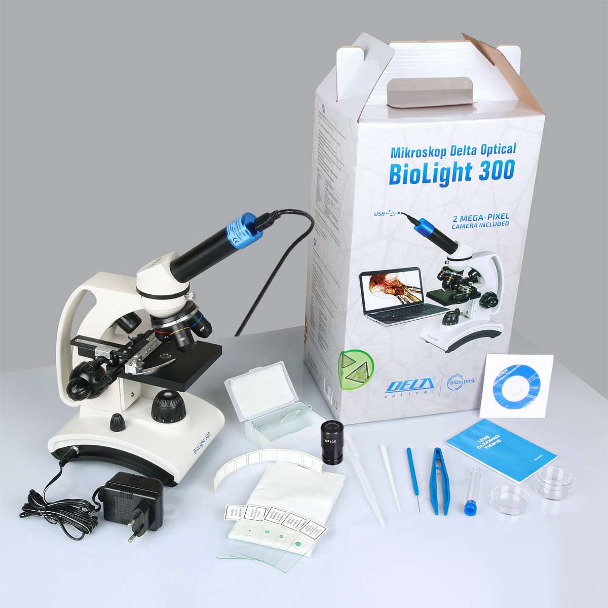
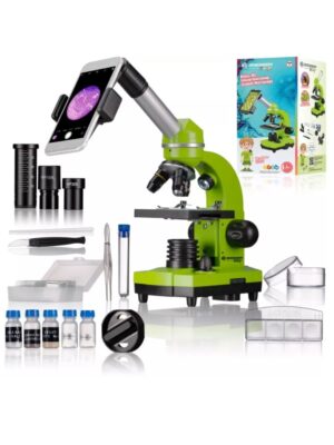
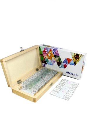
- the set includes a set of ready-made preparations (you can start observing immediately after unpacking), preparation tools (for preparing your own preparations) and materials for keeping the optics clean — a detailed list in the «equipment» tab
The Delta Optical BioLight 300 microscope enables the observation of simple biological preparations (plant and animal tissues), insects, minerals, plants, stamps, coins, precious stones, electronic circuits and much more.
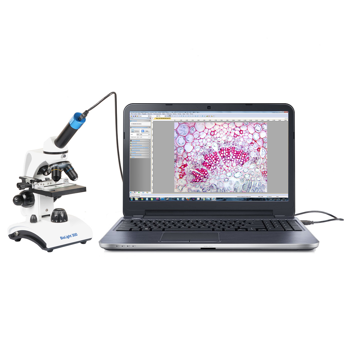
The camera is a very important element of the microscope equipment. The use of the camera in education and didactics in schools makes it possible to present the image on a larger screen, via a multimedia projector or an interactive board. Having a microscope with a camera at home makes children more likely to view the image on a computer screen, as they often find it difficult to focus on the image seen through the eyepiece. However, on the monitor, we can immediately indicate to the child interesting elements of the preparation.
Thanks to the excellent Delta Optical DLTCam Basic camera with a resolution of 2 million pixels used in conjunction with our microscope, you can prepare a multimedia presentation from hand-made preparations or enrich homework at school.
The microscopic exercises given in the manual will ensure long, fascinating hours of working with the microscope!
Note: for the microscope, it is recommended to use AA rechargeable batteries (fingers). The use of ordinary batteries is allowed, but only when the microscope is disconnected from the mains . Otherwise, it may damage the device or even fire.
There are various microscopes available on the market, offered with the so-called «Barlow lens», which further increases the magnification factor. Theoretically speaking, this is true, because the Barlow lens additionally enlarges the image transmitted through the lenses to the eyepiece (remember that the image resolution is determined by the lens, it results from the laws of physics). But a real Barlow lens is not a single glass element, as such a design solution causes chromatic aberration and spherical aberration (aberrations are image distortions in the optics). And most importantly: such magnification does not bring any new details to the microscopic image! A professional Barlow lens (such as in astronomical telescopes) consists of three or more optical elements,
A magnification of 400x is a sufficient approximation for a young explorer who will see the cellular structures of sections of preparations from plant and animal tissues, see unicellular organisms inhabiting the aquatic environment (e.g. protozoa, rotifers, algae, free-living nematodes). In reflected light, insects, fabrics, stamps and coins will be very visible. Students work with microscopes in primary and middle schools on the offered magnification. Thanks to our microscope, you will observe such experiments as, for example, the phenomenon of plasmolysis in the onion storage husk cells, or the effect of sucrose on yeast cells (Saccharomyces sp.).
Technical Specifications:
The most important functions of the Delta Optical DLT CamViewer software included with the camera:
— English, Russian and other languages version
— live image preview, with a choice of resolution
— image frozen, scaling, fit to the window, full-screen preview, vertical and horizontal mirroring for proper reproduction
— mode automatic time-lapse saving (available formats: * .bmp, * .dib, * .rle, * .jpg, * .jpe, * .jpeg, * .jif, * .jfif, * .png, * .tif, *. tiff, * .pcx, * .tga, * .jp2, * .j2k, * .tft)
— built-in viewer of saved images (available formats: * .avi, * .wmv)
— exposure time adjustment: automatic and manual
— balance adjustment white: automatic and manual (color temperature)
— manual color adjustment: hue, saturation, brightness, contrast, gamma
— work in color or monochrome mode and in negative mode
— length calibration function against a standard and saving calibration schemes
— functions for geometric measurements: angle, point, line, line parallel, two parallel lines, perpendicular lines, rectangle, ellipse, circle, circle inscribed in a circle (ring), two circles, arc, polygon — calculation of the area and perimeters of figures
— the possibility of carrying out measurements on recorded photos and live images
— the ability to export measurement results in text form to a spreadsheet or save on an image
— adding text annotations
— grids and rulers, reference scale that allows you to read the currently used magnification and scale
— creating and managing layers
— folding a stack of microscopic images saved in the Z axis into an image with an extended depth of field (EDF function)
— «stitching» function — combining microscopic images into a 2D panorama
— image processing through various filters and segmentation
function — automatic counting of objects in the image
Automatic installation on Windows 2000 / XP (SP2) / 2003 / Vista / 2008 / Windows 7/8 / 8.1 (32-bit and 64-bit), Linux and MacOS. The camera does not require drivers.
Camera technical specification:
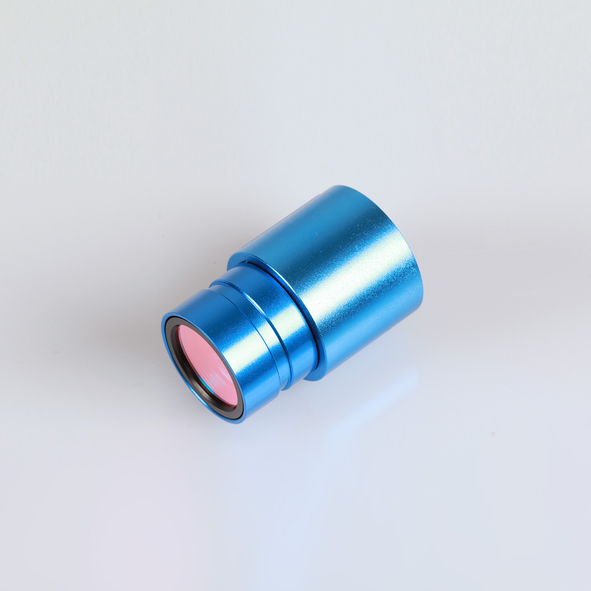
- Camera model name: Delta Optical DLT-Cam Basic 2 MP:
- digital color microscope camera
- maximum resolution: 1920×1080 pixels (2 megapixels)
- sensor size (diagonal): 1 / 2.8 «
- pixel size: 2.9 µm x 2.9 µm
- sensitivity: 1300 mV
- dynamic range: 73 dB
- analog-to-digital converter: 8-bit RGB
- signal-to-noise ratio: 43 dB
- frame rate (FPS): 38 fps for 1920×1080 pix; 38 fps for 960×540 pix
- assembly in tubes with an internal diameter of 23.2 mm
- interface: USB 2.0
- power supply: DC 5V via the USB interface of the computer
- included Polish-language Delta Optical DLT-CamViewer software with live image preview, photo and video recording, built-in functions
- image parameter adjustment, filters and measurement functions
- included USB cable for connection with a computer, 30 mm and 30.5 mm adapters




Отзывы
Отзывов пока нет.